Key points
- The uterine cavity is examined with a 2.9 mm telescope, with no incision.
- In the consulting room, no anaesthesia, no fasting, no hospital stay: 5 to 10 minutes.
- Best done in the first half of the cycle, roughly between day 6 and day 12.
- Targeted biopsy and minor procedures possible at the same time; result explained at the end.
Contents
What is diagnostic hysteroscopy?
The hysteroscope is a very thin telescope, 2.9 mm in diameter, connected to a camera. It is passed through the vagina and the cervix, with no incision. The cavity is opened with saline, which makes the examination far better tolerated than older techniques.
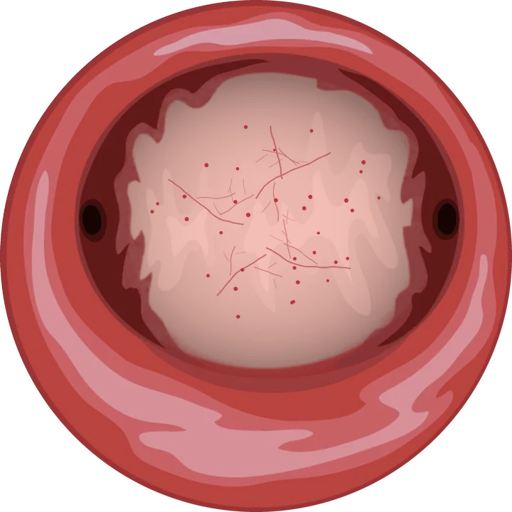
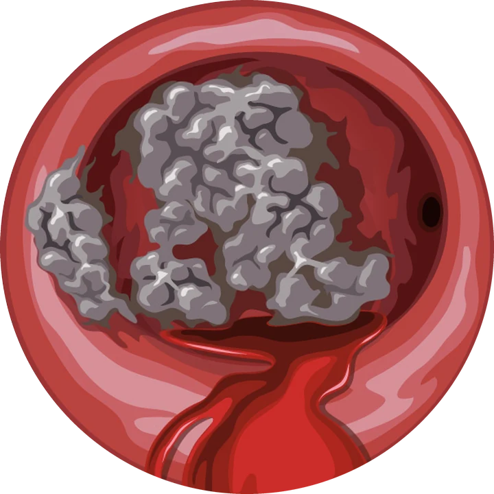
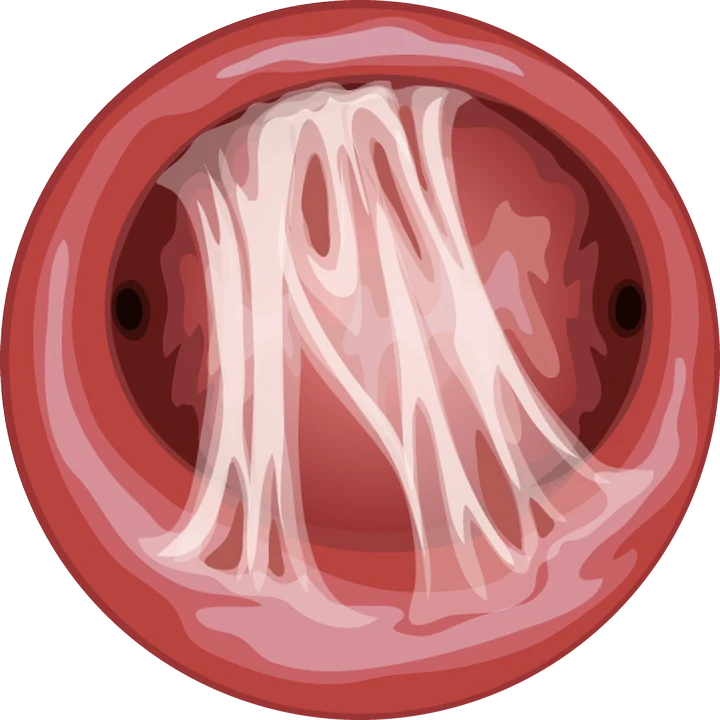
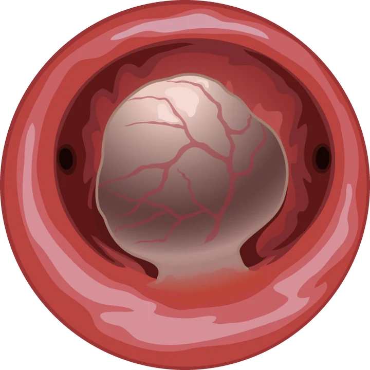
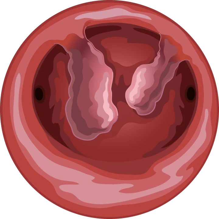
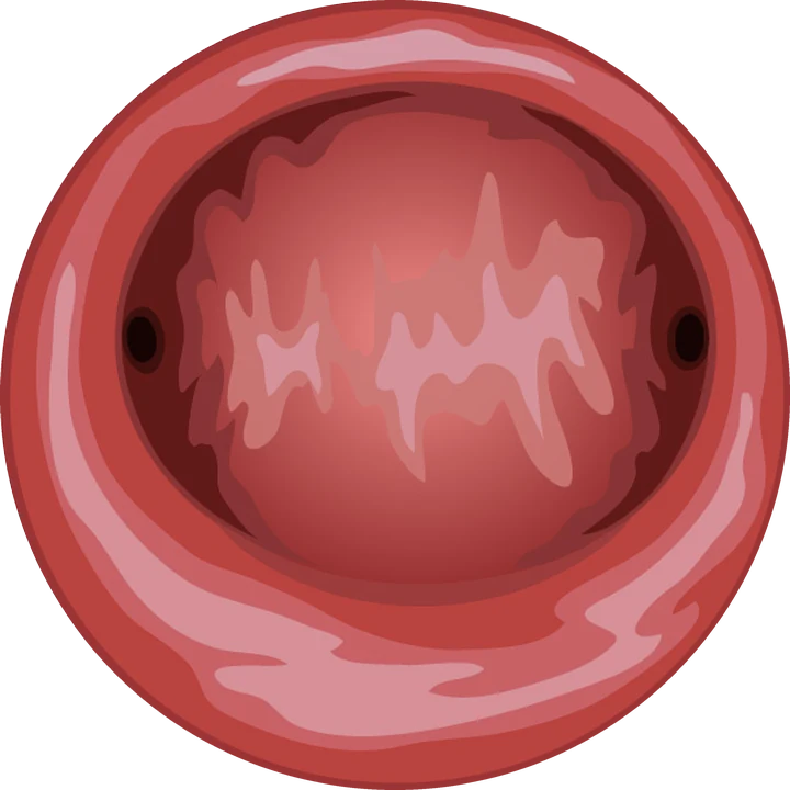
The cervical canal, the isthmus, the endometrium and both tubal openings are examined. Hysteroscopy confirms what the ultrasound suggests, allows a targeted biopsy and often treats a small lesion at the same time.
When is diagnostic hysteroscopy offered?
It is offered when an abnormality of the uterine cavity is suspected, usually after an ultrasound scan:
- heavy periods or bleeding between periods;
- any bleeding after the menopause, to rule out endometrial cancer;
- fertility assessment, before IVF or after implantation failure;
- recurrent miscarriage;
- suspected chronic endometritis;
- an IUD whose threads can no longer be seen.
Hysteroscopy is not routine: whether it is needed depends on your symptoms, the ultrasound and your plans.
How does the examination work?
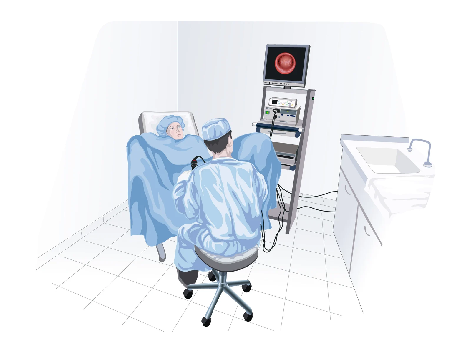
- Before. No heavy preparation, no fasting. A simple painkiller an hour before if you wish. Pregnancy test if in doubt.
- Positioning, 2 minutes. Gynaecological position. The speculum helps locate the cervix and is removed as soon as the telescope is inserted; no clamp is placed on the cervix. The screen faces you if you want to watch.
- Examination, 5 to 10 minutes. The telescope passes through the cervix into the cavity, opened with saline. Biopsy or minor procedure if needed.
- Afterwards. The result is explained and the report, with images, is handed to you.
Including the discussion and report, allow about 30 minutes. The most common sensation is a brief cramp, similar to period pain: see Is hysteroscopy painful?
When in the cycle?
In the first half of the cycle, after your period ends and before ovulation, roughly between day 6 and day 12. After the menopause or on continuous contraception, it can be done at any time.
After the examination
You can return to your activities the same day. Light spotting is possible for 24 to 48 hours. If a biopsy was taken, the result is available in about a week and is explained at a consultation, together with the next steps.
Seek advice promptly in case of fever, increasing pain or heavy bleeding.
Diagnostic or operative?
| Criterion | Diagnostic · clinic | Operative · hospital |
|---|---|---|
| Aim | See, biopsy, minor procedures | Remove or correct a lesion |
| Procedure time | 5 to 10 min | 15 to 45 min |
| Anaesthesia | None | General or regional |
| Fasting | No | Yes, as advised by the anaesthetist |
| Hospital stay | No | Day case, home the same day |
| Back to activity | Immediate | 24 to 48 h |
When a lesion needs removing and this cannot be done in the consulting room, it is treated by operative hysteroscopy, through the same natural route, as a day case.
Cover and fees
In France, diagnostic hysteroscopy is a listed CCAM procedure reimbursed by national health insurance. Dr Kadoch is contracted in sector 2 with free fees (not OPTAM), so extra fees may apply. The amount is given when booking. International patients should check with their insurer.
Frequently asked questions
Is diagnostic hysteroscopy painful?
With a speculum removed as soon as the telescope is in place, no clamp on the cervix and a 2.9 mm telescope, most women feel discomfort similar to period cramps, which stops when the examination ends. No anaesthesia is needed.
Do I need to fast?
No. Diagnostic hysteroscopy in the consulting room needs no fasting, no anaesthesia and no hospital stay.
How long does the examination take?
The examination itself takes 5 to 10 minutes. Including the discussion and report, allow about 30 minutes.
Can a polyp be removed during the examination?
Yes, a small polyp can often be removed at the same time in the consulting room with micro-instruments passed through the hysteroscope. Larger lesions are treated in theatre as a day case.
References
Written and reviewed by Dr Olivier Kadoch · 31 October 2026
This page is for information; it does not replace a consultation. Every situation is discussed individually.