Key points
- Malformations present from birth, classified by the ESHRE/ESGE consensus.
- A septate uterus is the most common; the septum may be partial or complete.
- 3D ultrasound is the key test; hysteroscopy shows the cavity and allows resection.
- Septum resection is no longer routine: it is decided according to each patient's history.
Contents
How are uterine malformations classified?
The uterus forms in the embryo when two ducts fuse and the wall between them is then absorbed. If either step stops, a malformation results. The European ESHRE/ESGE consensus classifies them as follows:
| Class | Shape | Example |
|---|---|---|
| U1 | Dysmorphic uterus | T-shaped uterus, including the DES uterus |
| U2 | Septate uterus | Partial or complete septum |
| U3 | Bicorporeal uterus | Bicornuate uterus |
| U4 | Hemi-uterus | Unicornuate uterus, with or without a rudimentary horn |
| U5 | Aplastic uterus | No functional uterus |

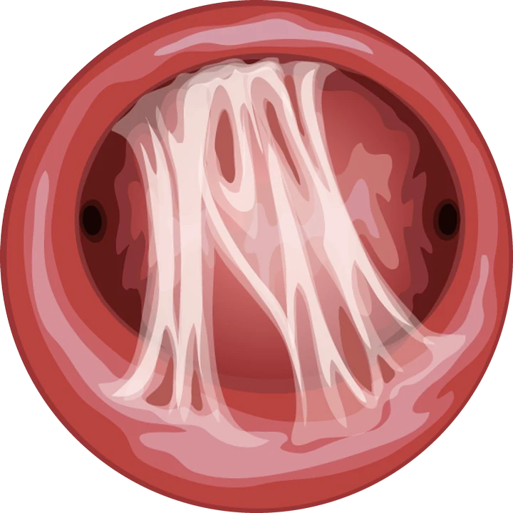
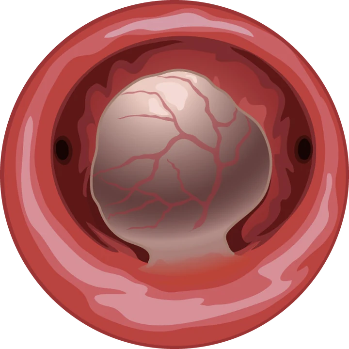
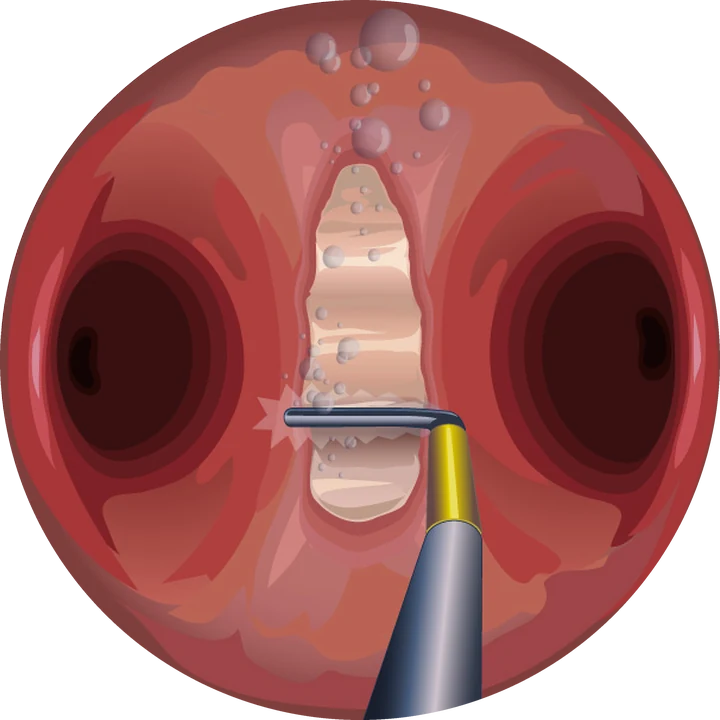
Hysteroscopic view · illustration
The septate uterus
A fibromuscular septum descends from the top of the uterus and divides the cavity in two, with a normal outer contour. It is often found during an assessment for miscarriage or infertility. European and American definitions of a septum differ, which explains differing opinions between centres.
Septum resection is done by hysteroscopy as a day case. Long offered widely, it is now debated: the randomised TRUST trial (2021) found no increase in live births after resection, and the ESHRE 2022 guideline no longer recommends it routinely. The decision is therefore made case by case, according to the size of the septum and your obstetric history.
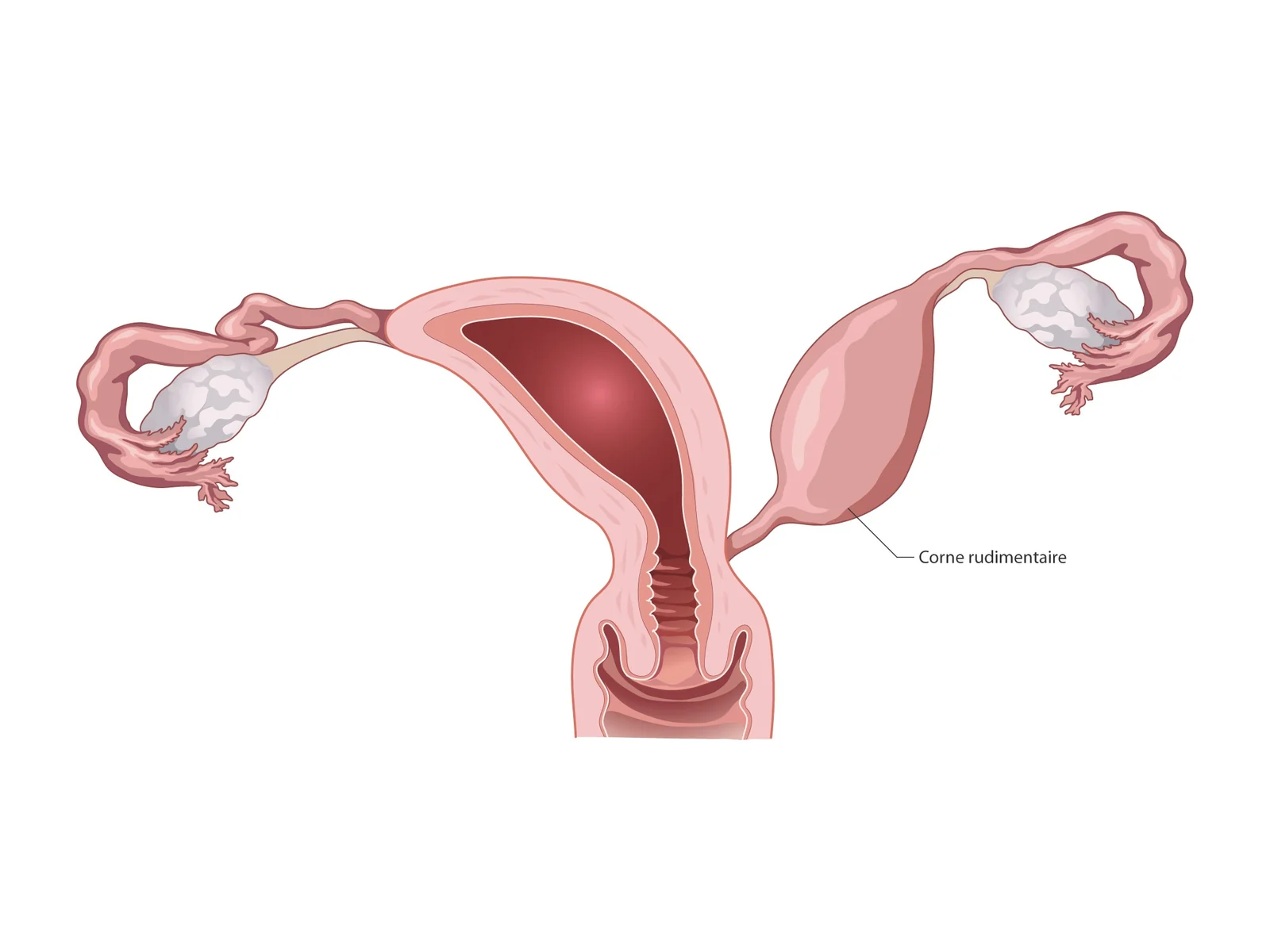
The unicornuate uterus
Only one half of the uterus has developed. The other half may persist as a rudimentary horn. If that horn contains endometrium, it can cause period pain, and a pregnancy can implant in it with a risk of rupture: laparoscopic removal is then recommended.
The DES uterus
Women exposed in utero to diethylstilbestrol (DES), prescribed in France until 1977, may have a T-shaped uterus with a narrow cavity. Their pregnancies are closely monitored, because of a higher risk of miscarriage, ectopic pregnancy and preterm birth.
How is the assessment done?
3D ultrasound shows both the cavity and the outer contour of the uterus, which distinguishes a septum from a bicornuate uterus without laparoscopy. MRI completes the picture if needed. Hysteroscopy shows the cavity from inside; it is done when a procedure is planned or to check the endometrium.
Frequently asked questions
Does a uterine septum prevent having a baby?
No. Many women with a septum have normal pregnancies. It can be associated with miscarriage or preterm birth, which justifies an assessment.
Should a uterine septum be operated on?
Not routinely. Since the TRUST trial and the ESHRE 2022 guideline, resection is decided case by case, according to the size of the septum and your obstetric history.
How do I know if my uterus is malformed?
3D ultrasound is the simplest and most accurate test. It is often done during an assessment for infertility or miscarriage.
What is a DES uterus?
A uterus altered by exposure to DES before birth, often T-shaped. It requires closer monitoring during pregnancy.
References
Written and reviewed by Dr Olivier Kadoch · 18 October 2026
This page is for information; it does not replace a consultation. Each situation is discussed individually.