Key points
- Often symptom-free; sometimes spotting, discharge or pelvic discomfort.
- At hysteroscopy: micropolyps, oedema and redness of the lining.
- Diagnosis rests on biopsy, looking for plasma cells (CD138 staining).
- Antibiotic treatment; its benefit on live births is still debated.
Contents
What is it?
It is a chronic inflammation of the endometrium, often linked to common bacteria. It is defined under the microscope by plasma cells in the supporting tissue of the lining. Its frequency varies widely between studies and methods: low in the general population, higher in women with implantation failure or recurrent miscarriage.

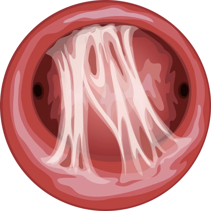
Hysteroscopic view · illustration
What hysteroscopy shows
| Sign | Appearance |
|---|---|
| Micropolyps | Tiny bumps under one millimetre, often numerous |
| Oedema | Thick, pale, irregular lining in the proliferative phase |
| Hyperaemia | Red areas, diffuse or patchy, sometimes with a white centre |
In a large series of more than 2,000 office hysteroscopies, these signs correlated well with biopsy. They remain suggestive rather than conclusive: the biopsy confirms.
Confirming the diagnosis
Endometrial biopsy is done in the consulting room, ideally in the first half of the cycle. CD138 immunohistochemical staining reveals the plasma cells. Bacteriological samples may be added.
Treatment and fertility
Treatment is with antibiotics, most often doxycycline for two weeks, adapted if a specific organism is found. A follow-up biopsy can confirm cure.
Several studies suggest better IVF outcomes after treatment in women with implantation failure. The European ESHRE guidelines, however, consider the evidence insufficient for routine screening: testing is offered case by case.
Frequently asked questions
Does chronic endometritis prevent pregnancy?
It is associated with implantation failure and miscarriage, but its exact role is still debated. It is mainly looked for in those situations.
How is it diagnosed?
Hysteroscopy shows suggestive signs; a biopsy with CD138 staining confirms.
How is it treated?
With antibiotics, usually for two weeks, sometimes followed by a check biopsy.
References
Written and reviewed by Dr Olivier Kadoch · 18 October 2026
This page is for information; it does not replace a consultation. Each situation is discussed individually.