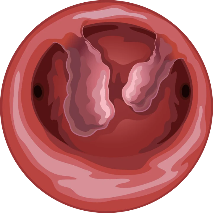
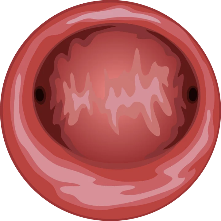
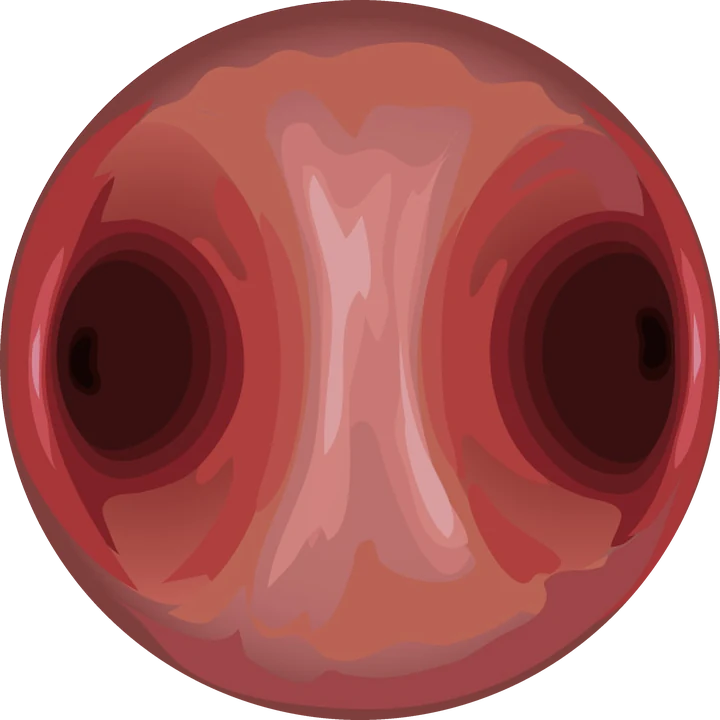
Key points
- A benign tumour of the uterine muscle, growing towards the cavity.
- Main symptom: heavy periods, sometimes with anaemia; possible infertility.
- The FIGO classification (types 0, 1, 2) determines the technique.
- Types 0 and 1: usually removed in one procedure; type 2: sometimes in two.
Contents
What is a submucosal fibroid?
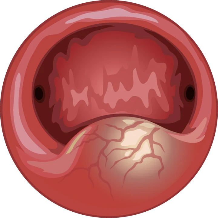
Fibroids (myomas) are very common benign tumours of the uterine muscle. They are classified by position: under the outer surface, within the muscle, or under the lining. A submucosal fibroid grows towards the cavity, in contact with the endometrium. It is the least common type but causes the most symptoms.
The FIGO classification describes how much of the fibroid lies within the cavity:
| FIGO type | Description | Hysteroscopic resection |
|---|---|---|
| Type 0 | Pedunculated, entirely within the cavity | One procedure |
| Type 1 | More than half within the cavity | Usually one procedure |
| Type 2 | Less than half within the cavity | Possible depending on size and muscle margin, sometimes in two stages |
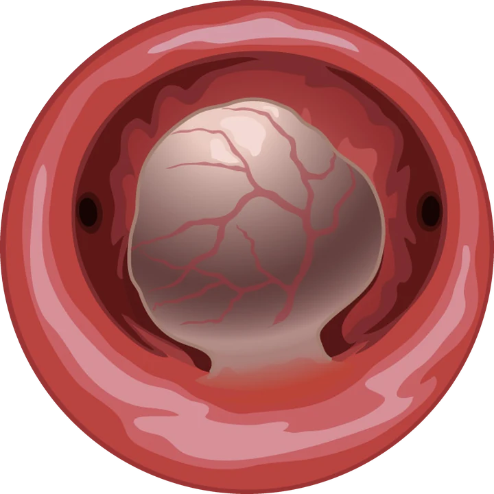
Hysteroscopic view · illustration
What are the symptoms?
- very heavy periods, with clots, flooding, double protection;
- fatigue due to iron-deficiency anaemia;
- bleeding between periods;
- infertility or miscarriage, as the fibroid distorts the cavity.
How is it diagnosed and the procedure planned?
Ultrasound, ideally 3D, locates the fibroid and measures the distance between the fibroid and the outer surface of the uterus. Saline infusion sonography and diagnostic hysteroscopy show how much lies within the cavity. MRI helps when there are several fibroids.
These findings estimate how difficult the resection will be (size, type, base, location) and tell you in advance whether a second stage is likely. Anaemia is corrected beforehand; a short hormonal treatment may be offered to shrink the fibroid or stop the bleeding.
How is hysteroscopic resection done?
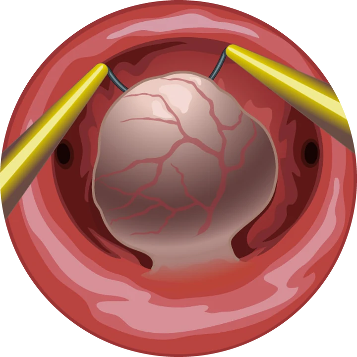
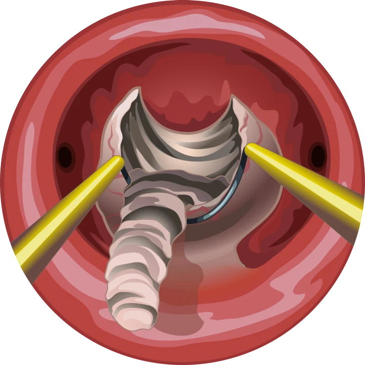
The procedure is a day case under general or regional anaesthesia. The fibroid is cut into small pieces with a bipolar resectoscope or a morcellator passed through the cervix, without any abdominal incision. The volume of distension fluid is measured continuously throughout, as part of standard safety rules.
The tissue removed is analysed. You go home the same day and resume activity within 24 to 48 hours.
What are the alternatives?
| Option | For whom | Limitation |
|---|---|---|
| Medical treatment | Moderate symptoms, approaching menopause, preparation for surgery | Effect lasts only while taken |
| Laparoscopic myomectomy | Mainly intramural or subserosal fibroids | Scar on the uterus |
| Embolisation | Multiple fibroids, no plans for pregnancy | Interventional radiology procedure |
| Hysterectomy | Other treatments have failed, family complete | Major surgery |
Ulipristal (Esmya) has not been used for this indication since 2020, because of serious liver injury.
What are the risks?
Complications are rare: bleeding, infection, perforation of the uterus, excess fluid absorption (prevented by monitoring the fluid balance), and adhesions in the cavity. A follow-up hysteroscopy may be offered after resection of a large fibroid, especially if you plan a pregnancy.
Frequently asked questions
Can a fibroid be removed without a scar?
Yes, when it is submucosal (FIGO types 0, 1 and often 2): it is removed by hysteroscopy, through the natural route.
How long does the procedure take?
Between 15 and 45 minutes depending on the size and type of the fibroid, as a day case.
Can the fibroid come back?
A removed fibroid does not regrow in the same place, but other fibroids can appear elsewhere in the uterus over time.
When can I try for a pregnancy?
Usually after one or two cycles, once the cavity has healed. The interval is confirmed after the procedure, depending on the depth of resection.
References
Written and reviewed by Dr Olivier Kadoch · 18 October 2026
This page is for information; it does not replace a consultation. Each situation is discussed individually.