Key points
- Always rule out pregnancy first.
- The cervix is examined and cervical screening should be up to date.
- An endometrial polyp is one of the commonest causes; it is removed by hysteroscopy.
- After a caesarean, brown spotting after periods suggests a caesarean niche.
Contents
The commonest causes
| Cause | Clues |
|---|---|
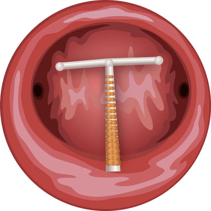
| Contraception | Low-dose pill, missed pills, implant or hormonal IUD, especially in the first months |
| Endometrial polyp | Irregular bleeding, sometimes after sex; seen on ultrasound and hysteroscopy |
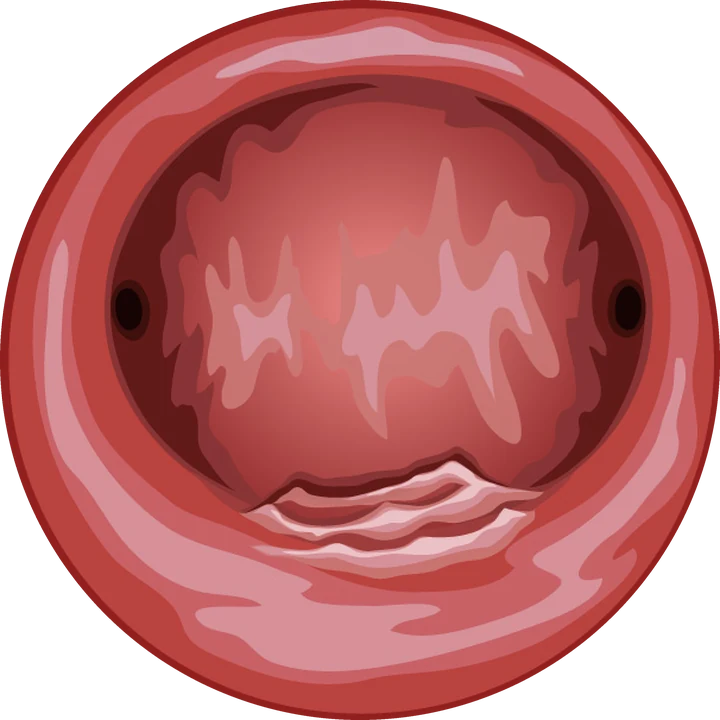
| Cervix | Ectropion, cervical polyp, infection or lesion: bleeding after sex |
| Infection | Chlamydia or other infection: discharge, pain, bleeding |
| Caesarean niche | After a caesarean: brown bleeding a few days after the period ends |
| Pregnancy | Pregnancy test in every case |
| Endometrial lesion | Rare before 45, to be ruled out with risk factors |
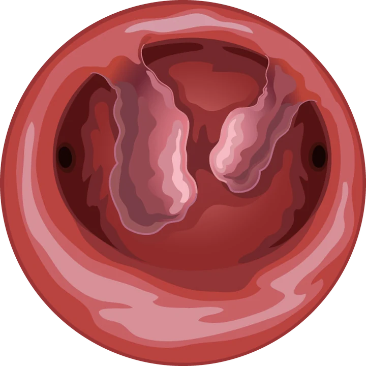
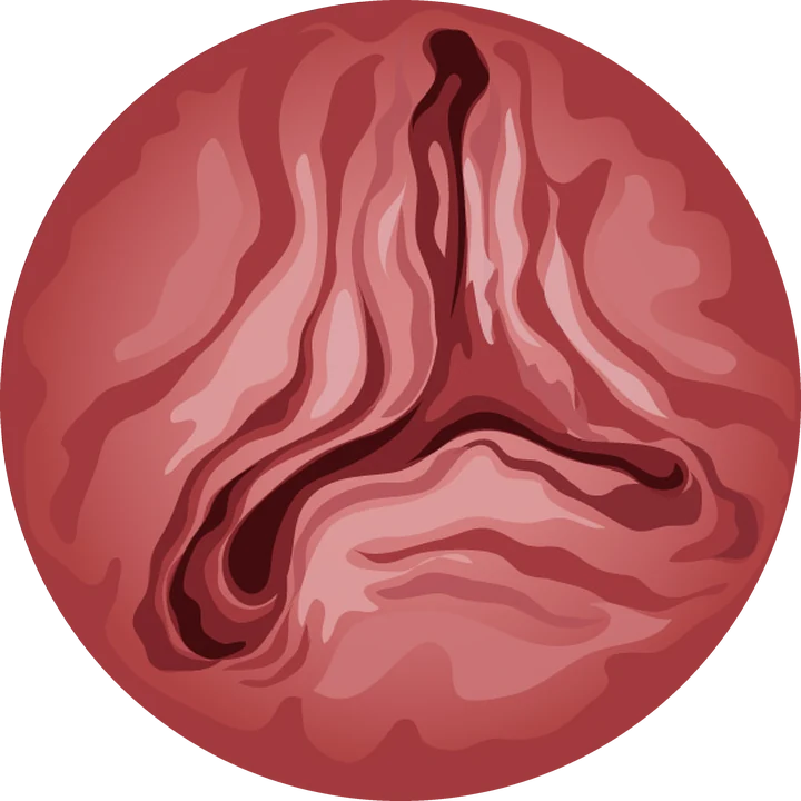
Hysteroscopic view · illustration
What work-up?
The work-up starts with a pregnancy test, a cervical examination and a check that cervical screening (smear or HPV test, depending on age) is up to date. An infection screen is done if needed. Pelvic ultrasound looks for a polyp, fibroid or caesarean niche.
Diagnostic hysteroscopy is offered when ultrasound shows an image in the cavity, when bleeding persists without a cause, or for a targeted biopsy. It also examines the cervical canal and the caesarean scar.
Treating the cause
- Polyp: hysteroscopic removal, often in the consulting room, with microscopic analysis;
- Contraception: change of method or dose;
- Infection: antibiotics, and partner treatment if needed;
- Cervical lesion: management based on screening and colposcopy results;
- Troublesome caesarean niche: hysteroscopic isthmoplasty when the wall is thick enough.
When to seek care quickly
Seek care promptly for bleeding in pregnancy, heavy bleeding, pain or fever, repeated bleeding after sex, or bleeding after the menopause.
Frequently asked questions
Is bleeding between periods serious?
Usually not: the cause is generally benign. It still warrants a work-up, especially if it persists or happens after sex.
Can a polyp cause bleeding between periods?
Yes, it is one of the commonest causes. It is removed by hysteroscopy, often in the consulting room.
Why do I have brown spotting after periods since my caesarean?
This may be a caesarean niche, a pouch in the scar where blood collects. Ultrasound and hysteroscopy show it.
References
Written and reviewed by Dr Olivier Kadoch · 18 October 2026
This page is for information; it does not replace a consultation. Each situation is discussed individually.