Key points
- Useful when ultrasound shows an abnormality, after implantation failure, or after a curettage or caesarean section.
- Not routine before a first IVF cycle: two large randomised trials showed no benefit in that setting.
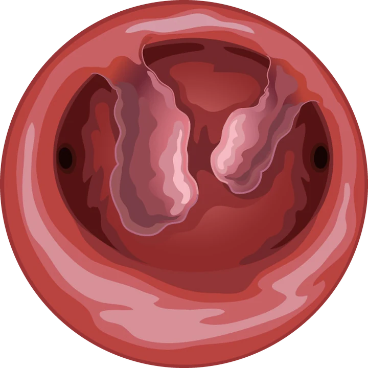
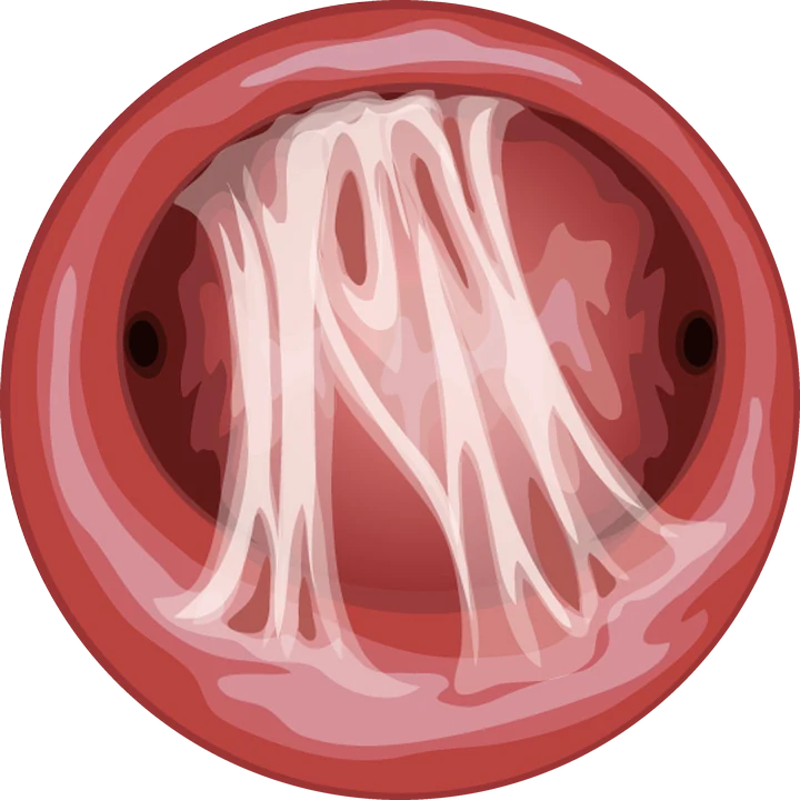
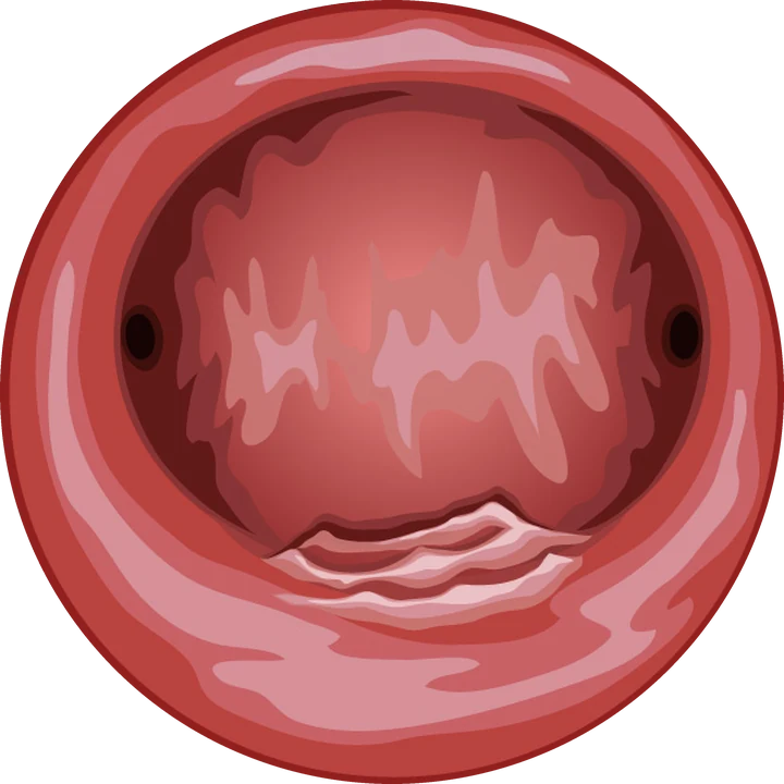
- Lesions most often corrected: polyps, submucosal fibroids, adhesions, caesarean niche.
- Endometrial scratching is not recommended; hysteroscopy does not assess tubal patency.
Contents
Why look inside the uterus?
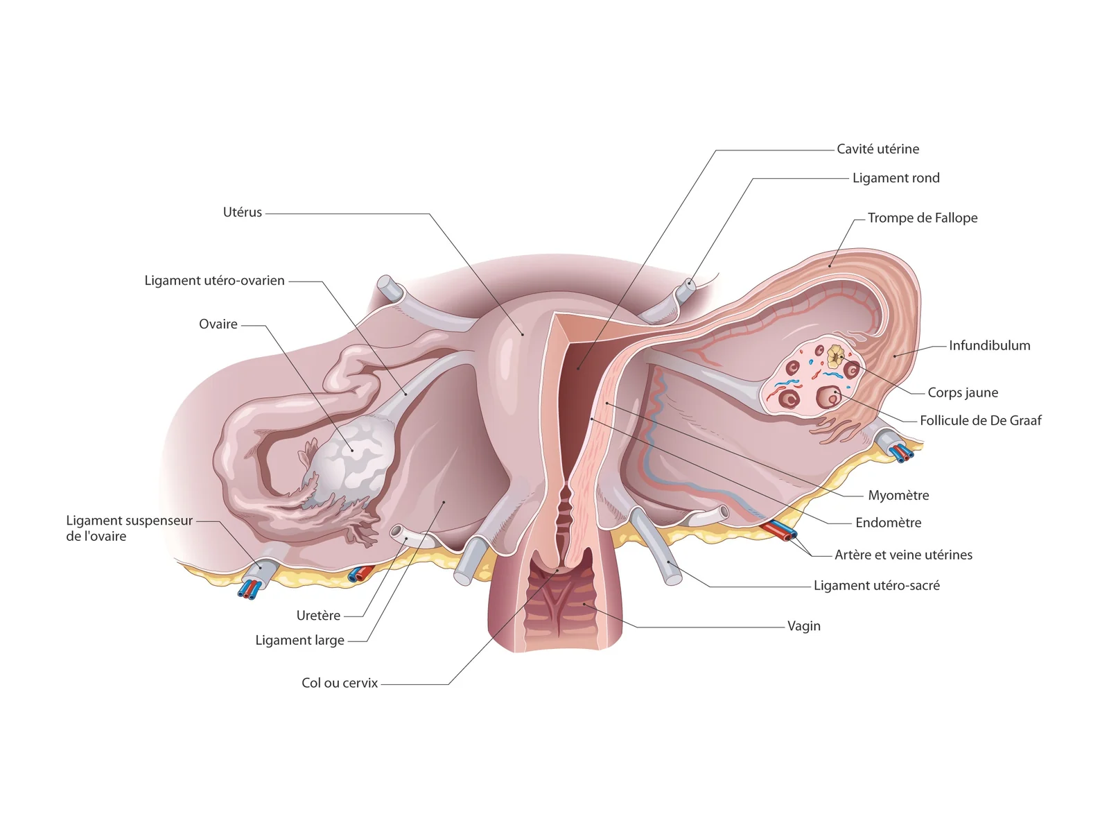
A polyp, a fibroid distorting the cavity, adhesions or a pouch in a caesarean scar can prevent an embryo from implanting or increase the risk of miscarriage. These lesions are common in women seen for infertility, and many cause no symptoms.
Hysteroscopy shows them directly and often treats them at the same time, without any incision.
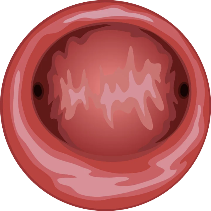
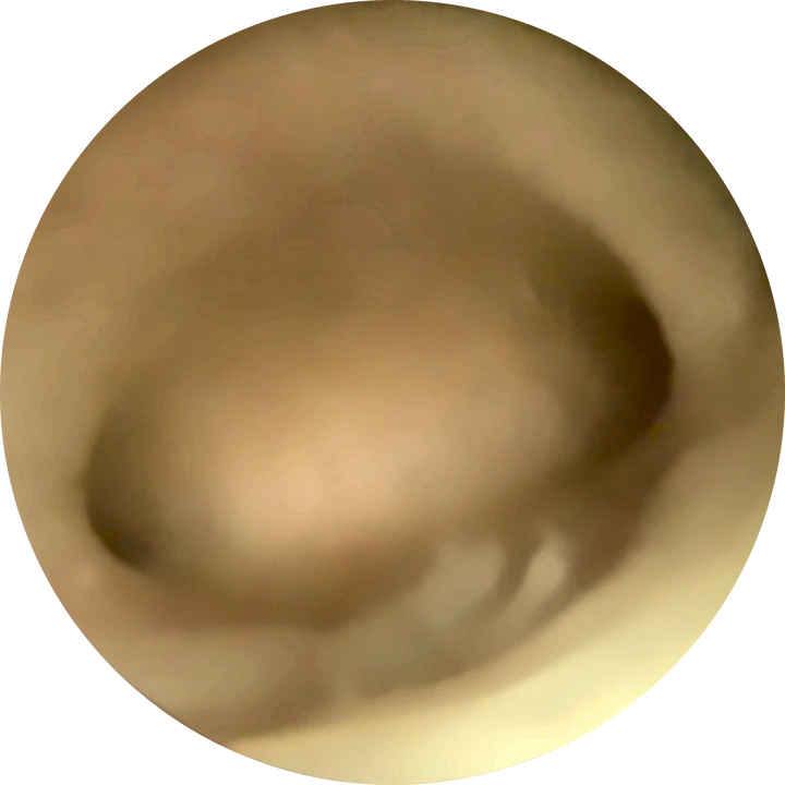
Hysteroscopic view · illustration
When is hysteroscopy useful?
| Situation | Value of hysteroscopy |
|---|---|
| Abnormal ultrasound image | Confirm and treat the lesion before the cycle |
| Recurrent implantation failure | Look for a missed lesion; part of the ESHRE work-up |
| Previous curettage or retained tissue | Look for adhesions |
| Previous caesarean and spotting after periods | Look for a caesarean niche |
| Recurrent miscarriage | Look for a malformation or adhesions, with 3D ultrasound |
| First IVF, normal ultrasound | No proven benefit as a routine test |
What does the evidence show?
Two randomised trials published in The Lancet in 2016 tested routine hysteroscopy. The inSIGHT trial, before a first IVF cycle, and the TROPHY trial, after failed IVF, found no increase in live births among women with a normal ultrasound. The 2019 Cochrane review reached the same conclusion.
When a lesion is present, however, correcting it matters. In a randomised trial, removing polyps before insemination markedly increased pregnancy rates. Hysteroscopy is therefore offered selectively, not to everyone.
Which lesions are corrected?
- Polyps: removed, often in the consulting room;
- Submucosal fibroids (FIGO types 0 to 2): hysteroscopic resection;
- Adhesions: division, then a follow-up check;
- Caesarean niche: isthmoplasty if the remaining wall is thick enough;
- Uterine septum: decided case by case since the TRUST trial.
Signs of chronic endometritis (micropolyps, oedema, redness) can also be looked for, with a biopsy; their role in IVF care is still debated.
What is not recommended
Endometrial scratching, offered to help implantation, showed no benefit in a large randomised trial and is not recommended by ESHRE. Hysteroscopy does not assess tubal patency either: that requires hysterosalpingography or contrast ultrasound.
How is it scheduled?
Diagnostic hysteroscopy is done in the first half of the cycle, before stimulation, in coordination with your fertility clinic. After a minor procedure in the consulting room, treatment can often start the next cycle. After a larger resection, one to two cycles is usual, sometimes with a follow-up hysteroscopy.
Frequently asked questions
Should every woman have a hysteroscopy before IVF?
No. With a normal ultrasound and a first IVF cycle, two large randomised trials showed no benefit. It is useful with an abnormal image, implantation failure, or a history of curettage or caesarean section.
Does hysteroscopy improve pregnancy rates?
Yes when it corrects a lesion such as a polyp or adhesions. When the cavity is normal, it does not change pregnancy rates.
How long between hysteroscopy and IVF?
Often the next cycle after a minor procedure in the consulting room; one to two cycles after a larger resection.
Does hysteroscopy check the fallopian tubes?
No. It shows the uterine cavity and the tubal openings, not whether the tubes are open; that requires hysterosalpingography or contrast ultrasound.
References
Written and reviewed by Dr Olivier Kadoch · 18 October 2026
This page is for information; it does not replace a consultation. Each situation is discussed individually.